How To Read Mri Brain
Mri brain is a specialist investigation that is used for the assessment of a number of neurological conditions.
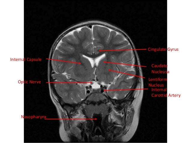
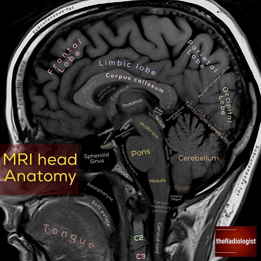
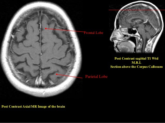
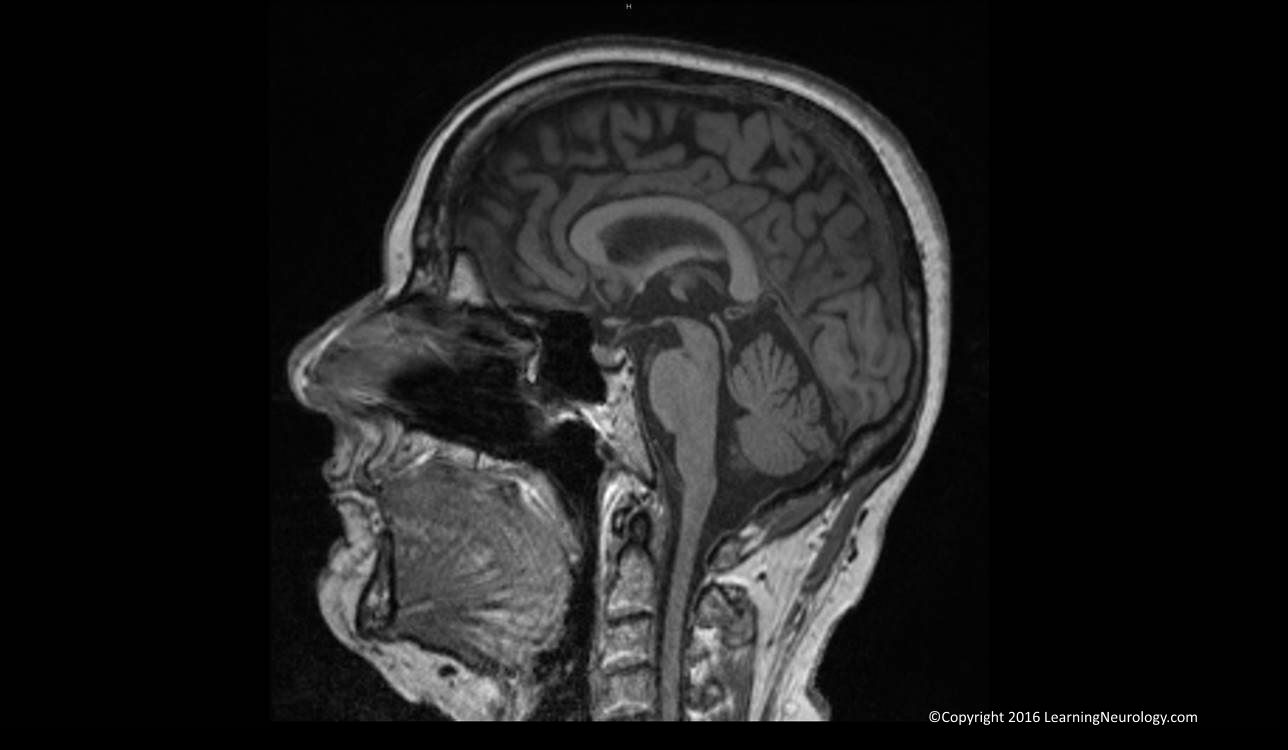
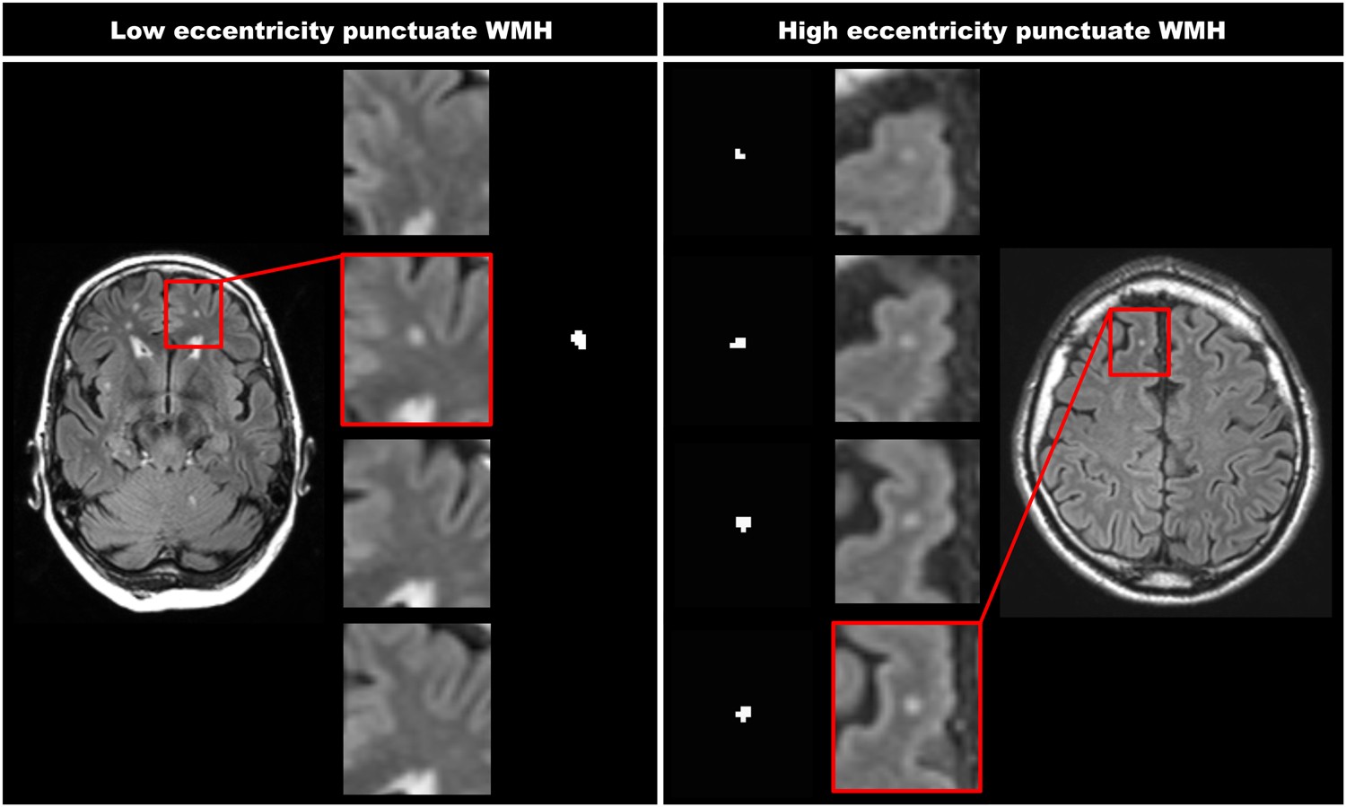
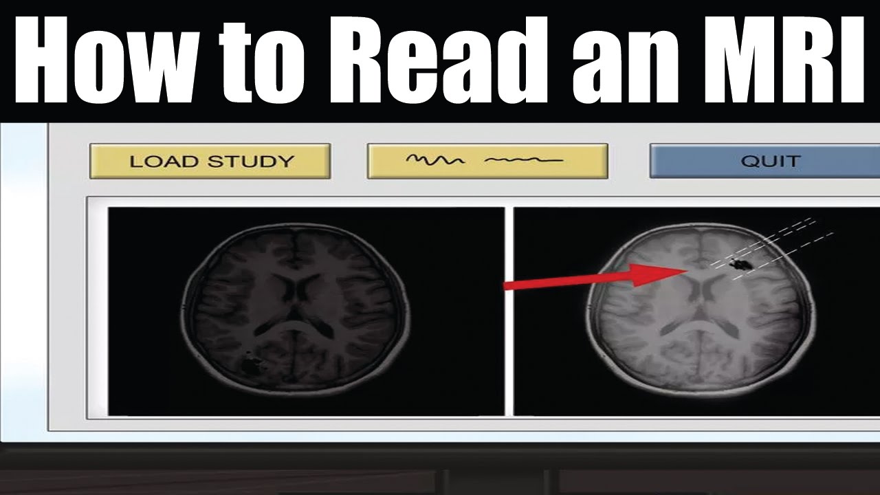
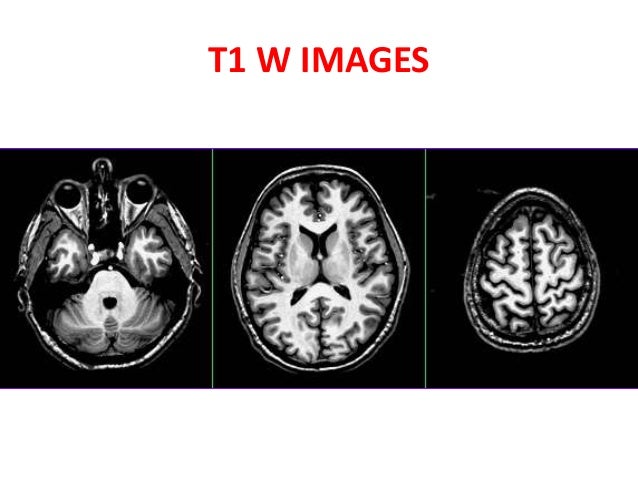
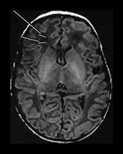
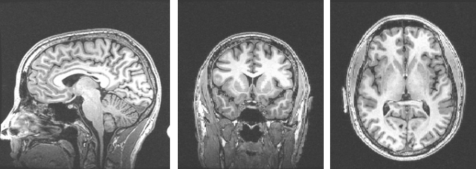
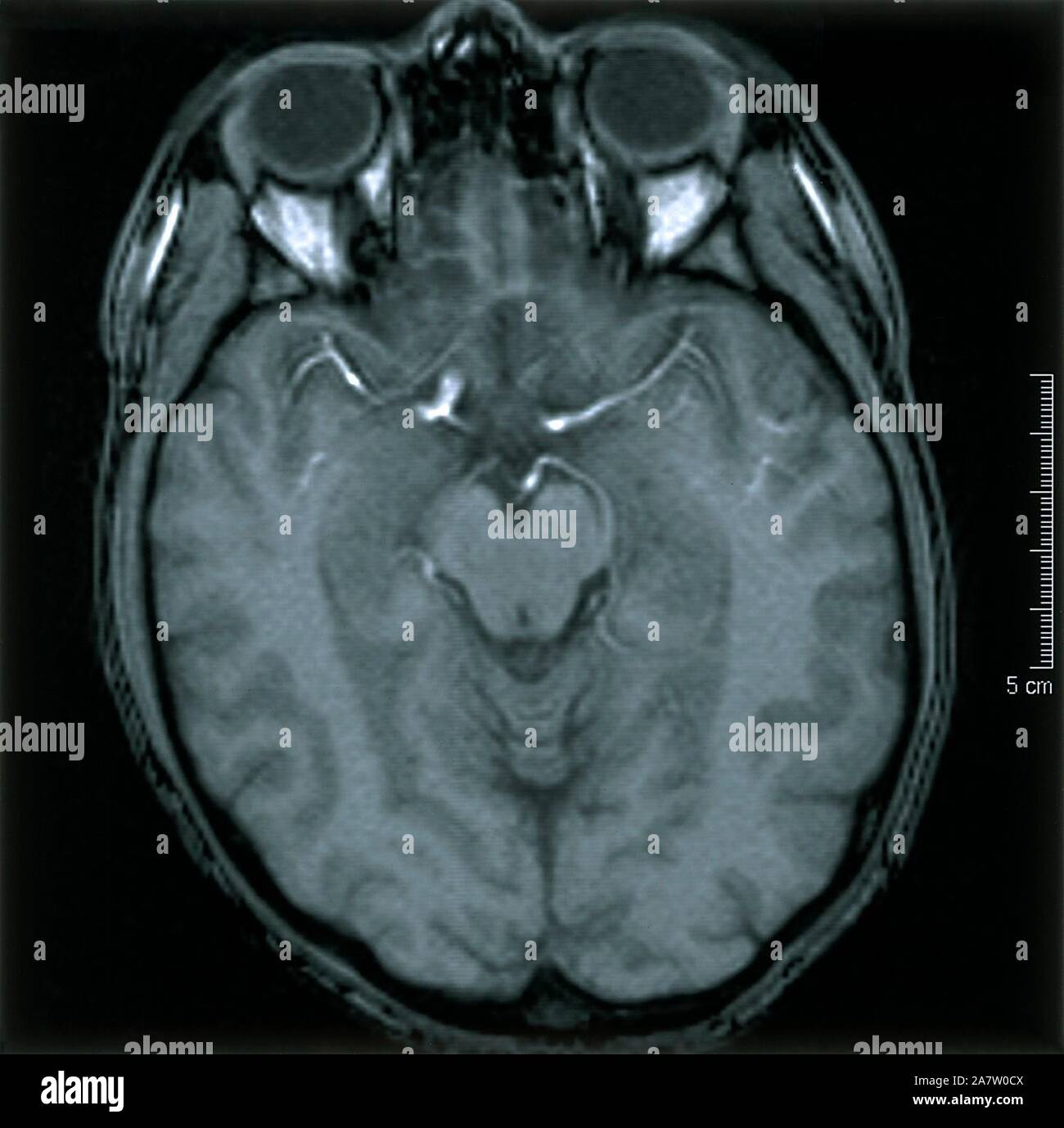
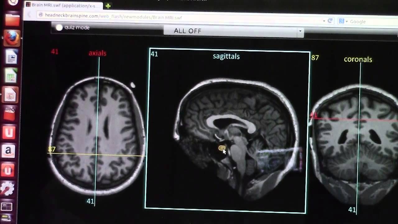
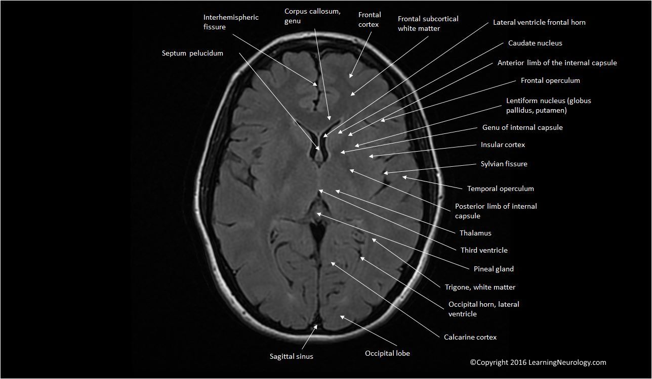
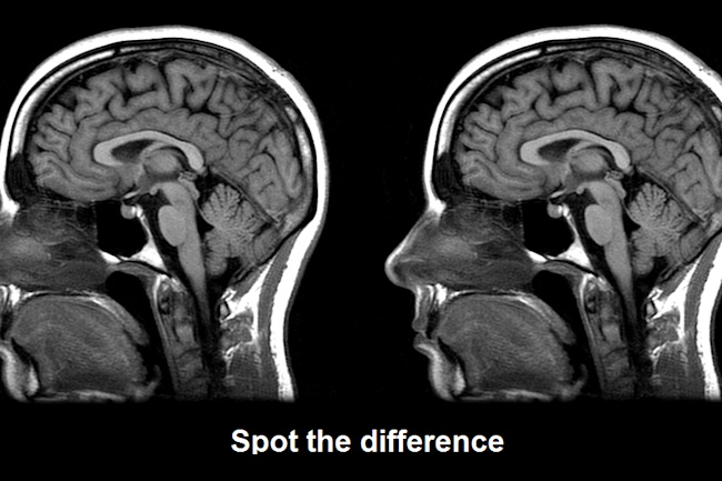
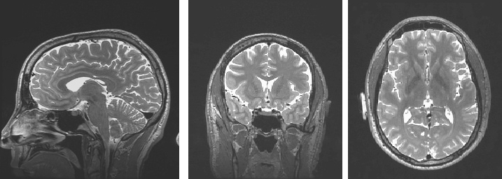
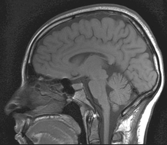
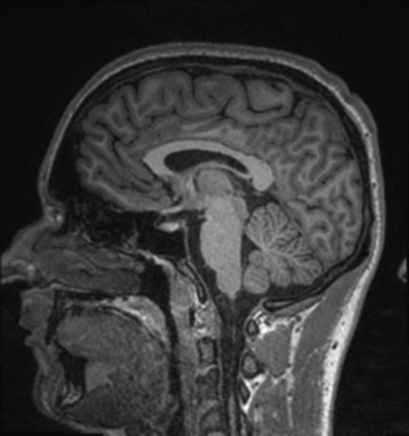
How to read mri brain. Normal mri brain in mid sagittal plane t1 with structures labeled. If you see small preview pictures of your mri images in the toolbar double click on the image you want to view. A brain lesion appears as a dark or light spot that does not look like normal brain tissues. Thus the brain mri analysis shall start from the ventricles going to the surrounding subcortical structures brain lobes cerebral cortex to the meninges and skull.
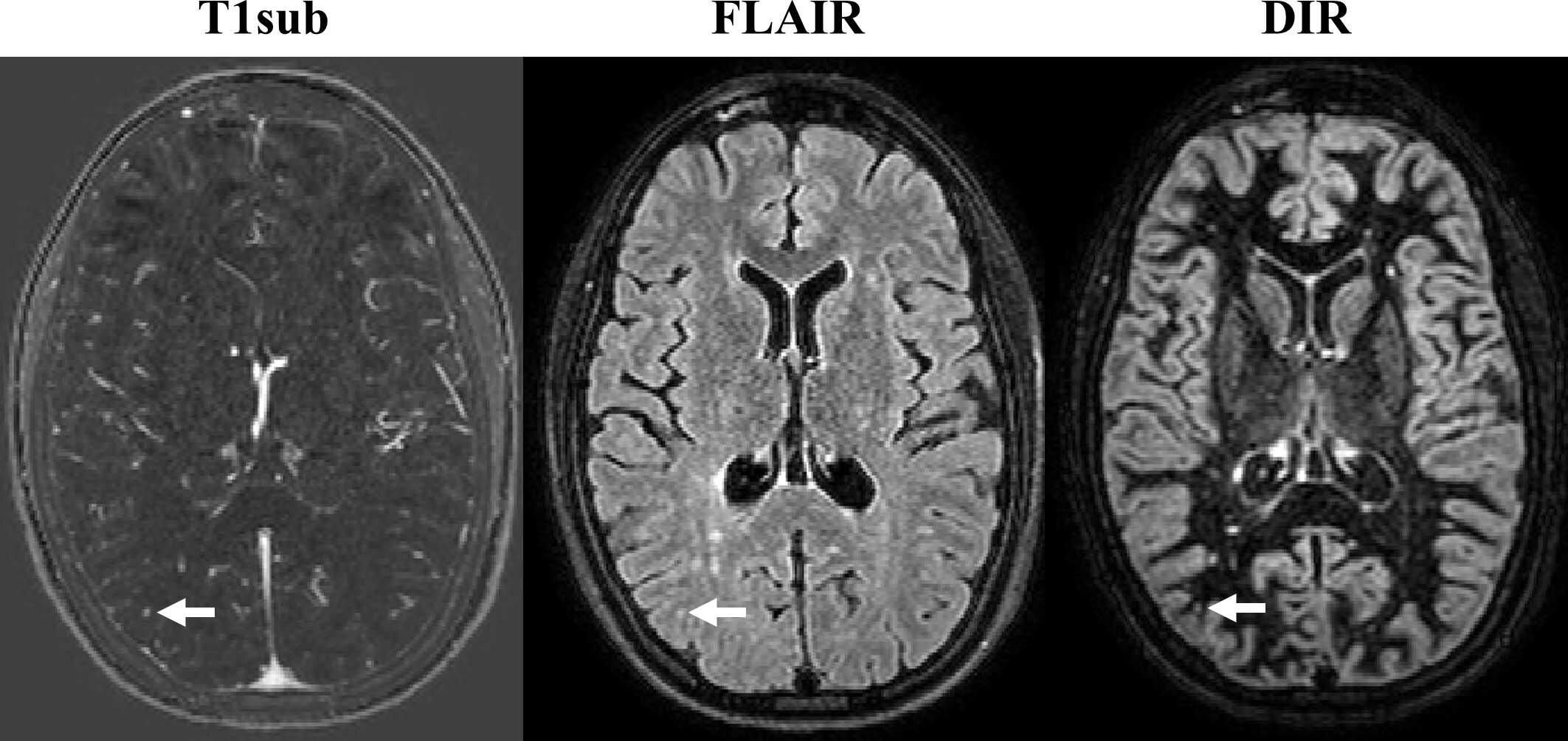
How to read a brain mri in this post lets take a look at the basics of how to read a brain mri because it often cant wait until the radiologist gets in in the morning. Add gadolinium contrast to evaluate tumor and abscess. The length of the procedure varies depending on the situation. Brain lesions may be present due to multiple sclerosis or as a result of an infection or a tumor.
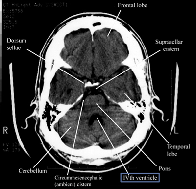
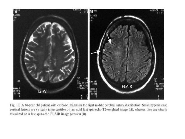
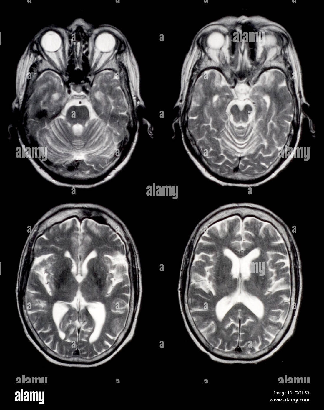
Observe the normal and abnormal structures. By looking at mri images your doctor can see details of blood flow and fluids surrounding the brain which can help determine abnormalities in the brain relating to arteries and veins. First acute ischemic stroke ais. It is the main method to investigate conditions such as multiple sclerosis and headaches and used to characterize strokes and space occupying lesions.
In general a brain mri will enable your doctor to examine blood flow and. A wide range of different mri images can be produced to help answer specific clinical questions. How to read brain mri brain mri examination should follow a systematic approach starting from the midline and going laterally. An mri scan is painless and noninvasive.
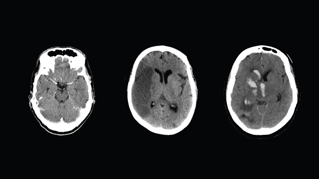
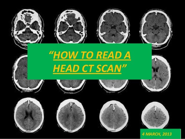
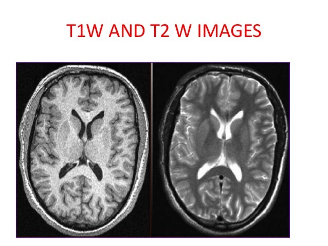
Look at all image planes available and compare the fat sensitive images with the water sensitive images. There are important safety issues regarding the use of mri. For brain hemorrhage however ct is the go to study. Image and patient information.
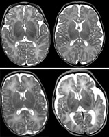
Most mri programs start with a large black space on one side of the screen and a smaller toolbar on the other side. In this article we take a close look at head mri scans in adults and children. An mri brain scan also shows brain lesions. A systematic approach is required for image interpretation.
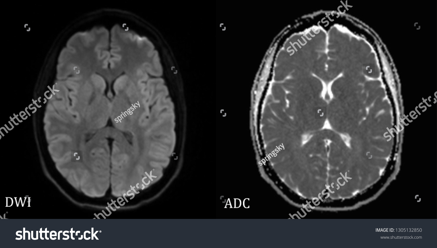
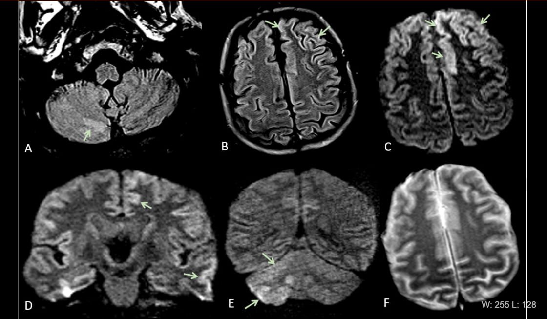
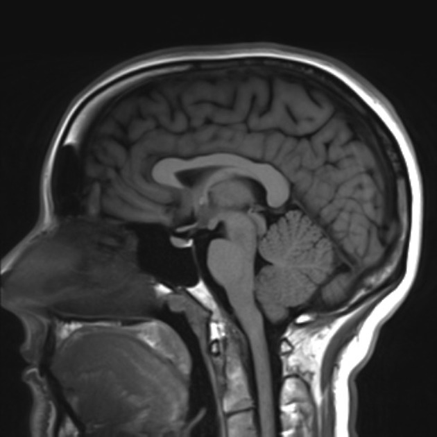
Sagittal mri brain t1 with blank labels. In this tutorial you will learn the basic knowledge required for mri image interpretation. An mri will show the stroke as bright signal on the diffusion weighted images and dark on the diffusion adc sequence. Approach to the structures sagittal.
An mri is the study of choice for tumor multiple sclerosis and ischemic stroke. Then look laterally at the parasagittal planes. When interpreting an imaging investigation always check the image and patient details. Any abnormality found should be considered with reference to the clinical question in mind.
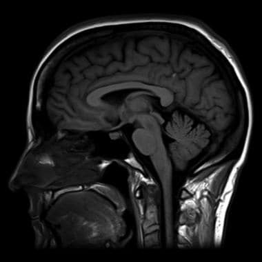
Sagittal mri brain t1. The 2 series you will want to pull up next to each other are the dwi diffusion weighted image adc apparent diffusion coefficient.
Normal Mri Brain

Differentiating Multiple Sclerosis Mimics On Mri Neurology Advisor
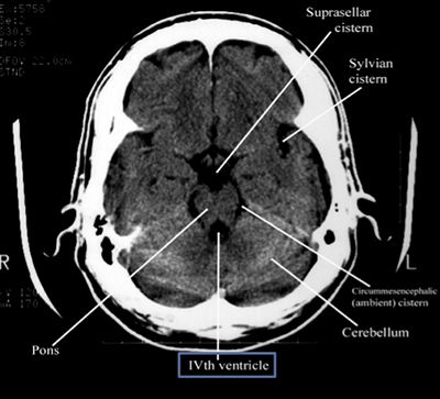
How To Read A Head Ct Emergency Medicine Newyork Presbyterian

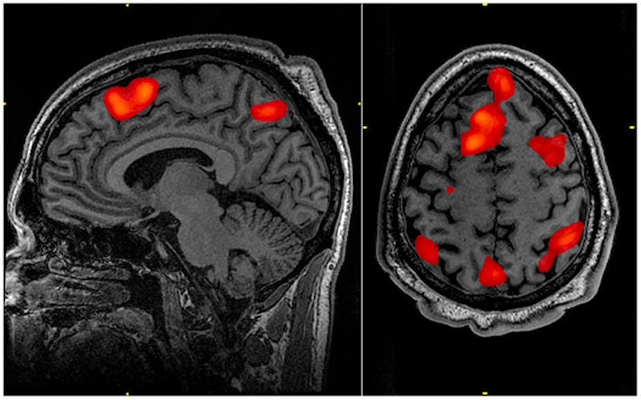
Children S Brains Reorganize After Epilepsy Surgery To Retain

Normal And Abnormal Mri Image Download Scientific Diagram