How To Read An Ultrasound At 5 Weeks
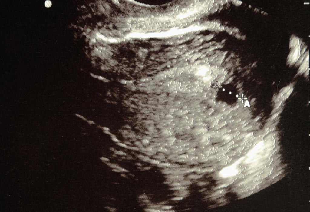
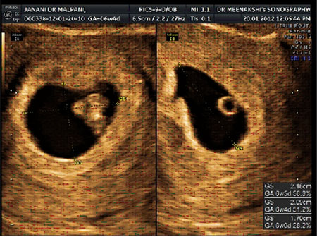
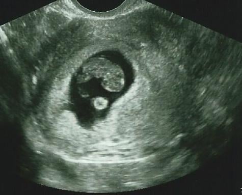
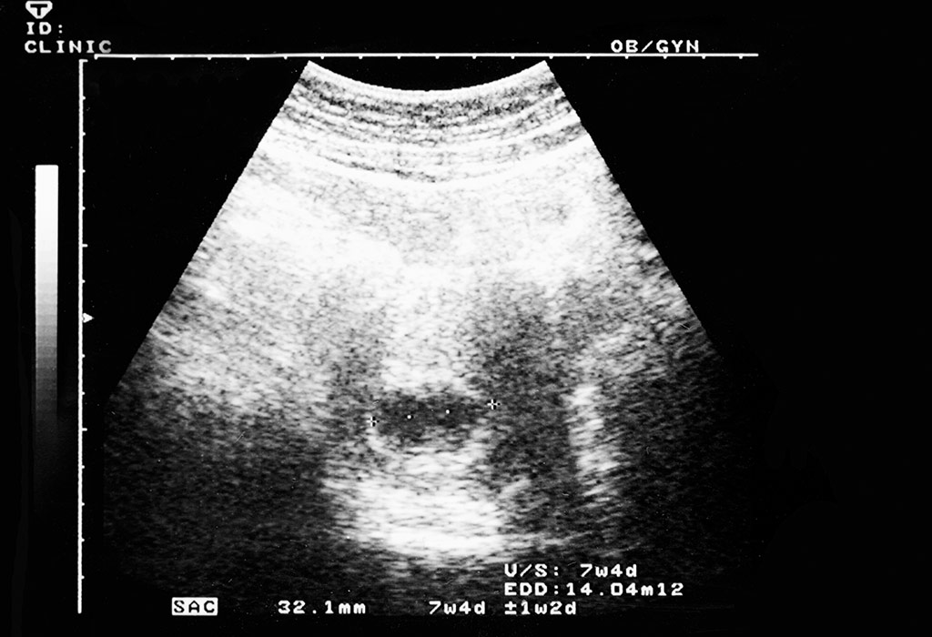
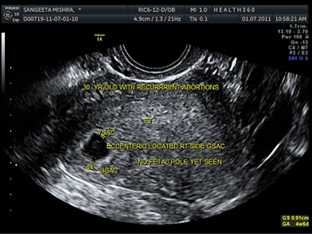
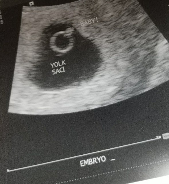
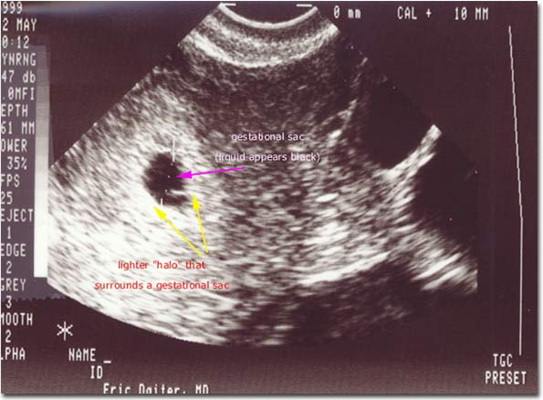
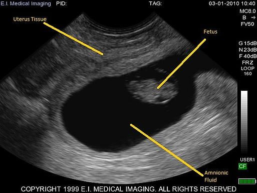
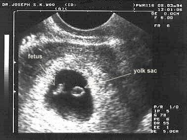
The ultrasound typically shows a gestational sac and within it we can see a 3 5 mm bubble like structure which is the yolk sac.

How to read an ultrasound at 5 weeks. Ultrasound is a non invasive immediate tool used to image tissue. For example if a woman has a positive pregnancy test the day of her missed period followed by confirmation with hcg blood test results and an ultrasound four weeks later shows a pregnancy at only five weeks gestational age the doctor may conclude that the woman has had a missed miscarriage. The difference will be astonishing. The positive pregnancy test a month earlier would indicate that the pregnancy should be more developed.
Various body tissues conduct sound differently. The yolk sac is first visible at 5 weeks and it is always present by 5 weeks and 4 days. This is usually about five and a half weeks after a pregnant womans last period. This is usually at four to five weeks after a pregnant womans last period.
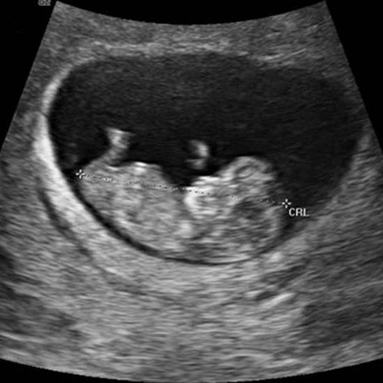
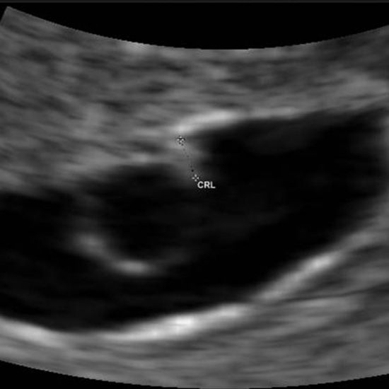
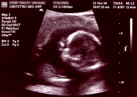
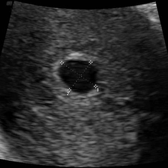
However at 12 weeks you should be able to see the head of your baby. Based on whether the twins are fraternal or identical you will be able to see them. Since the connecting stalk is short the embryonic pole is found near the wall. I just came back from my first ultrasound with this pregnancy and at 5 weeks 5 days ive a 12 mm mean sac measurement is 9mm gestational sac but nothing else no pole or yolk sac.
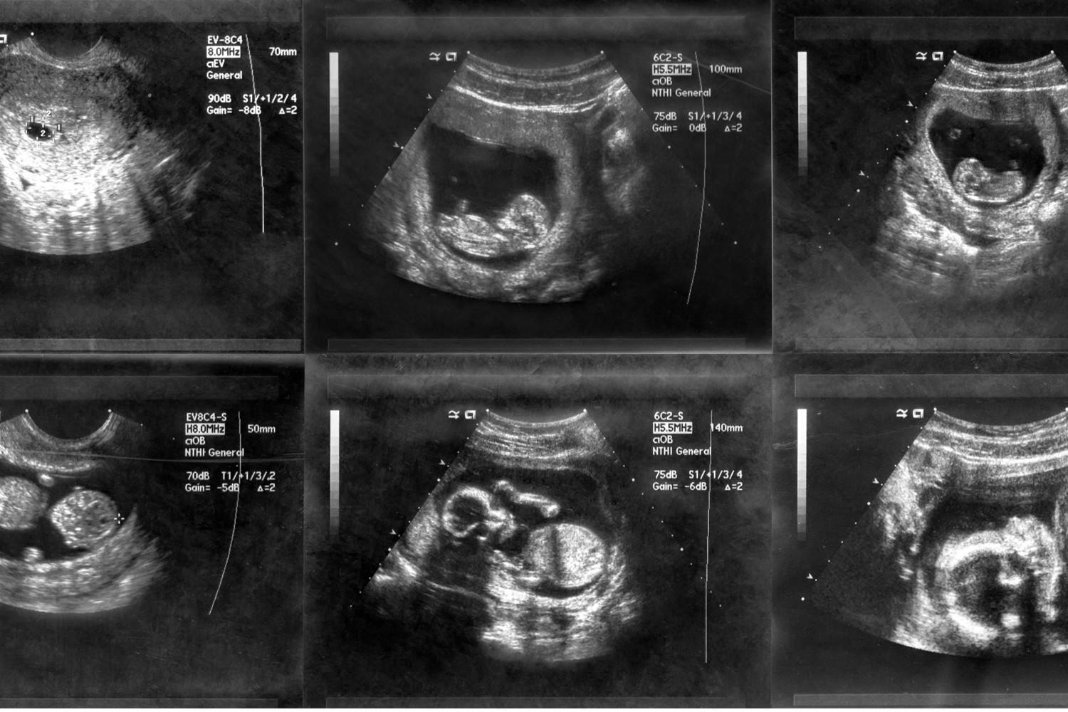
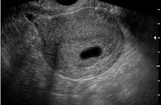
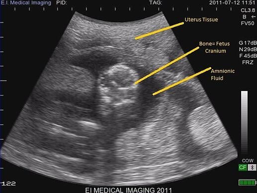
The ultrasound commonly shows a small collection of fluid within the lining of the uterus that represents the early development of the gestational sac. Im a little concerned as it was empty but the doc didnt seem worried and said that it was quite usual not to see anything at this point. It will not penetrate bone like an x ray. To read an ultrasound picture look for white spots on the image to see solid tissues like bones and dark spots on the image to see fluid filled tissues like the amniotic fluid in the uterus.
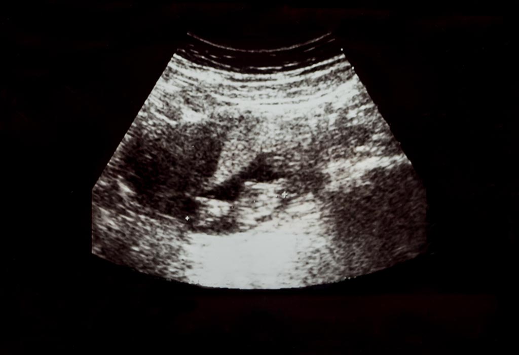
Not every woman will undergo an ultrasound at 5 weeks of pregnancy. So the first step to help you read the ultrasound image is to be familiar with the anatomy that you are imaging. This will involve a wand that will be inserted internally and images will appear on a screen. On your 5 weeks pregnant ultrasound you should be able to see your gestational sac and the yolk sac which is always present when you are 5 weeks pregnant.
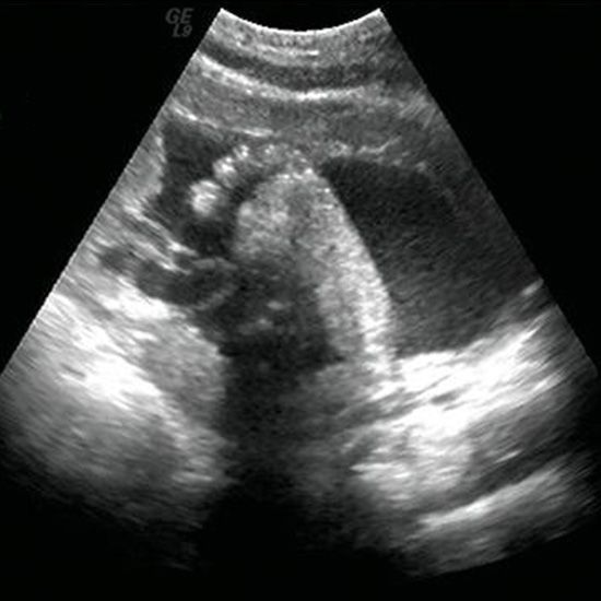
If she does it will be conducted via a transvaginal ultrasound. If you are trying to read an ultrasound at 20 weeks. Some tissues absorb sound waves while others reflect them. The embryonic pole appears adjacent to the yolk sac soon showing cardiac activity.
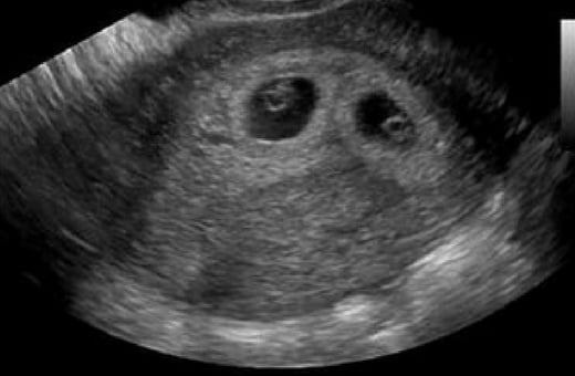
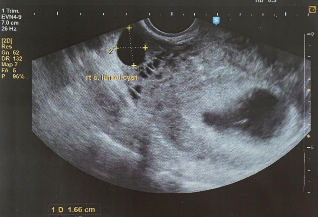
There are lacunary structures cavities or spaces at the site of implantation. Yes by performing an ultrasound scan at week 5 your doctor or the technician will be able to tell whether you are carrying twins or not.



:max_bytes(150000):strip_icc()/pregnant-couple-having-sonogram-186366123-5bff1a2cc9e77c005114eb76.jpg)

/female-nurse-performing-ultrasound-on-pregnant-woman-in-examination-room-697537711-5bff19cfc9e77c0051c989fc.jpg)

/doctor-using-ultrasound-on-pregnant-woman-591405835-595195b43df78cae81c346ba.jpg)





:max_bytes(150000):strip_icc()/ultrasound-showed-no-gestational-sac-2371356-color-V3-8136d007eb8e4306a7b876517e3638d9.png)







/cdn.vox-cdn.com/uploads/chorus_image/image/64568322/1748810.0.jpg)















/twin-ultrasound-106005791-5b8d9f79c9e77c002ca1159b.jpg)


